PYCAD BLOG

Insights on Medical Imaging, AI & DICOM

Guides, project breakdowns, and lessons learned from building medical imaging platforms — written by the PYCAD team.

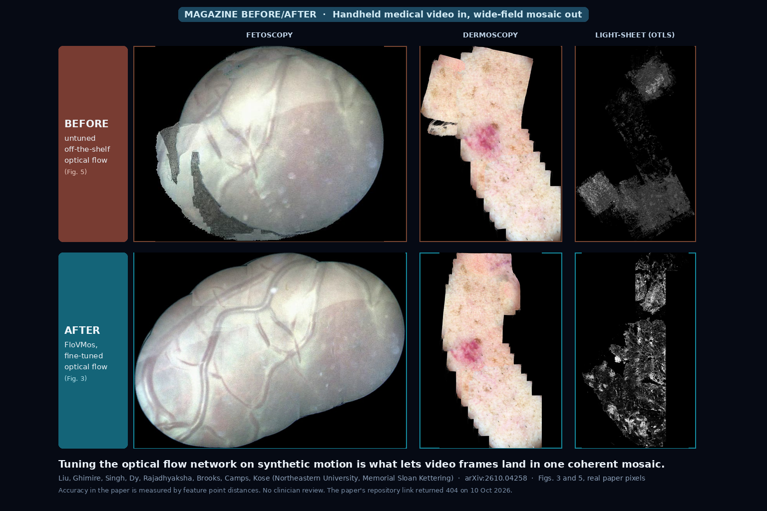
Before and after mosaics of fetoscopy, dermoscopy and light-sheet microscopy video: the top row, stitched with an untuned optical flow model, shows grey patches and broken pieces; the bottom row, from the fine-tuned FloVMos model, is continuous (Liu et al., arXiv:2610.04258, Figs. 3 and 5)

Handheld Endoscope or Microscope Video In, One Wide-Field Tissue Mosaic Out at About Ten Frames per Second From a Modality-Tuned Optical Flow Model

Feed FloVMos video from a handheld endoscope, dermoscope or confocal microscope and it stitches the frames into one growing wide-field mosaic, at about ten frames per second on 1 megapixel frames in the authors’ research code. The trick is fine-tuning an optical flow network per device on synthetic motion, so no hand-labelled flow is needed. Several real test sets are one or two videos, accuracy is a feature distance check, a GPU is required, and the repository linked in the paper returned a 404 when I checked. Liu et al., arXiv:2610.04258.
Read More »
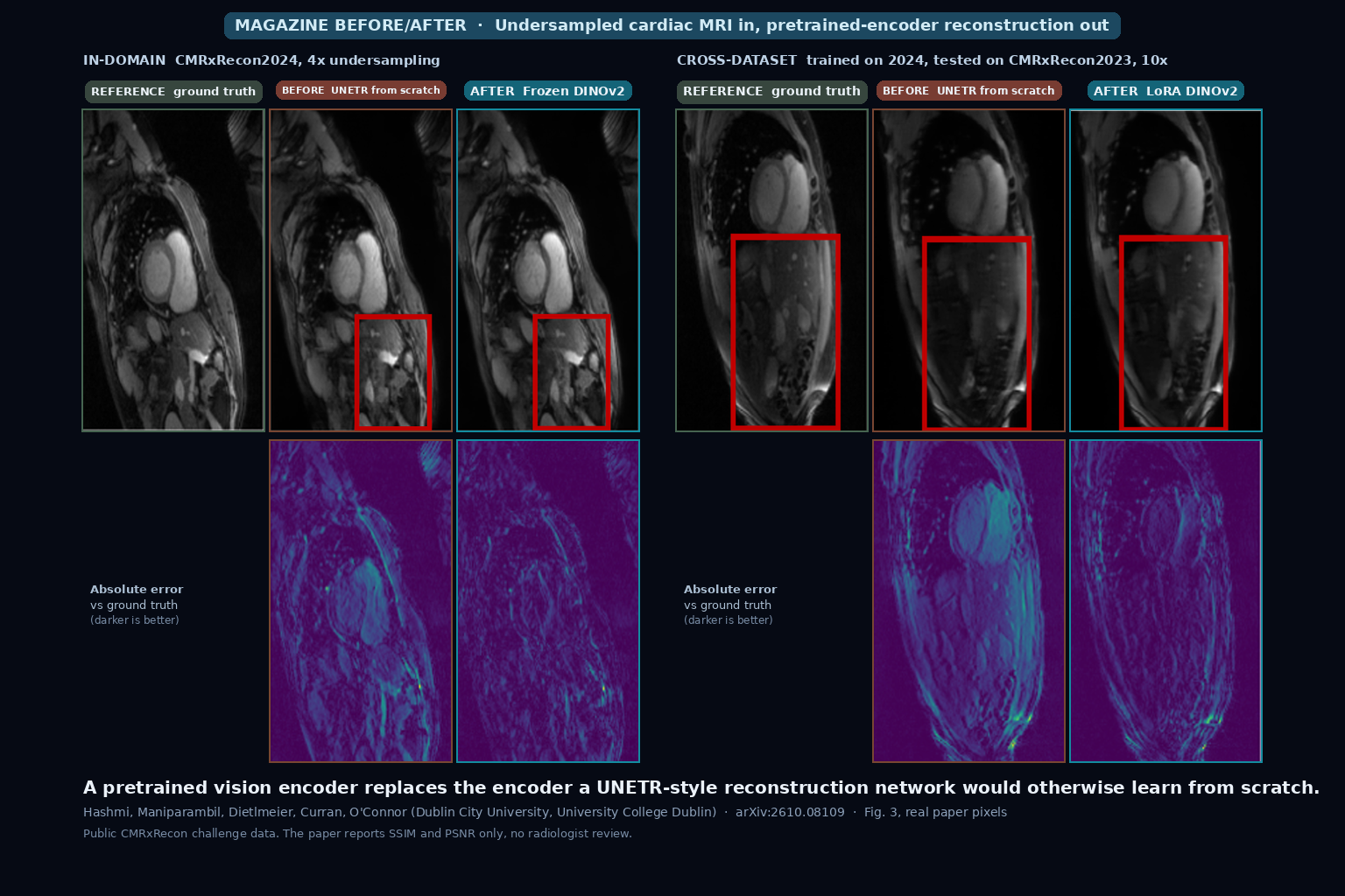
Two panels of cardiac MRI slices with absolute error maps below, each showing the ground truth, a UNETR trained from scratch, and a reconstruction from a pretrained DINOv2 encoder, at 4x acceleration in-domain and 10x acceleration across datasets (Hashmi et al., arXiv:2610.08109, Fig. 3)

Undersampled Cardiac MRI In, Sharper Reconstruction Out From a Frozen Pretrained Vision Encoder With About Half the Trainable Weights

Hand an undersampled cardiac MRI slice to a UNETR-style network whose encoder is a frozen pretrained CLIP, BiomedCLIP or DINOv2 model, and the reconstruction beats the same network trained from scratch, with about 49 percent fewer trainable parameters. The gap holds with 5 percent of the training data and across two challenge datasets. It is a 2D study on public CMRxRecon data scored with SSIM and PSNR only, with no reader study, and the linked GitHub repository held only a README when I checked. Hashmi et al., arXiv:2610.08109.

Read More »
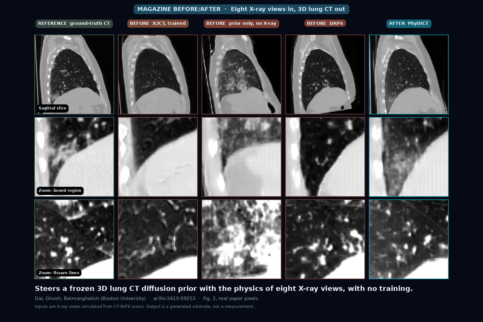
Three rows of lung CT crops, a sagittal slice and two zooms, comparing the ground-truth CT, X2CT, the diffusion prior alone, DAPS, and PhyDiCT reconstructed from eight X-ray projections (Dai et al., arXiv:2610.09253, Fig. 2)

Eight X-ray Projections In, a Generated 3D Lung CT Volume Out With No Training but About 22 Minutes of GPU Time

Give it eight simulated X-ray projections around the chest and it steers a frozen, text-conditioned lung CT diffusion model until its own rendered X-rays match, returning a 3D volume with no paired training. It takes about 22 minutes per volume on an RTX 6000, is built for lung CT only, and was tested on X-rays simulated from CT-RATE scans, so the output is a plausible estimate, not a measurement. Apache 2.0 code. Dai et al., arXiv:2610.09253.

Read More »

We build custom medical imaging platforms — advanced DICOM viewers, AI segmentation, and the clinical systems around them.

Copyright © 2026 PYCAD. All Rights Reserved.