Generic selectors
Exact matches only
Search in title
Search in content
Post Type Selectors
PYCAD BLOG

Insights on Medical Imaging, AI & DICOM

Guides, project breakdowns, and lessons learned from building medical imaging platforms — written by the PYCAD team.

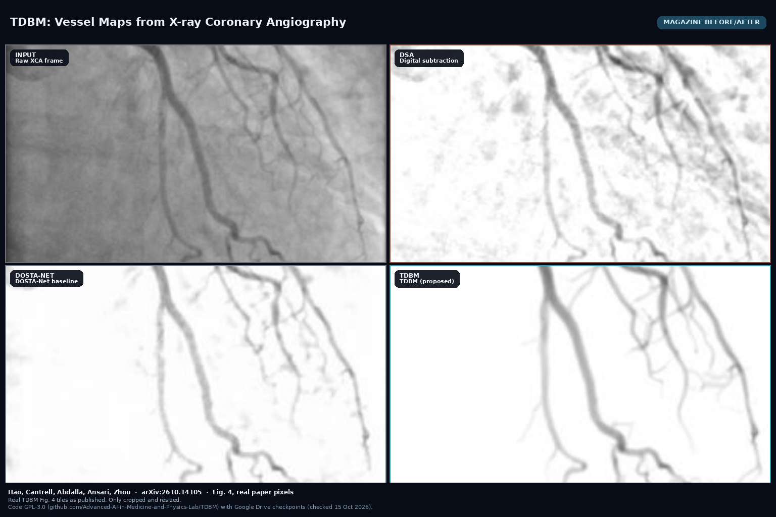
Four panels from Fig. 4: raw XCA input, DSA, DOSTA-Net, and TDBM vessel map on the same internal coronary angiography frame (Hao et al., arXiv:2610.14105)

TDBM: Vessel Maps from X-ray Coronary Angiography

TDBM, by Hao, Cantrell, Abdalla, Ansari and Zhou (arXiv:2610.14105), recovers contrast-preserving coronary vessels from X-ray angiography sequences with a temporal diffusion bridge trained on physics-informed synthetic data, without manual vessel labels. Code is GPL-3.0 on GitHub with Google Drive checkpoints.

Read More »
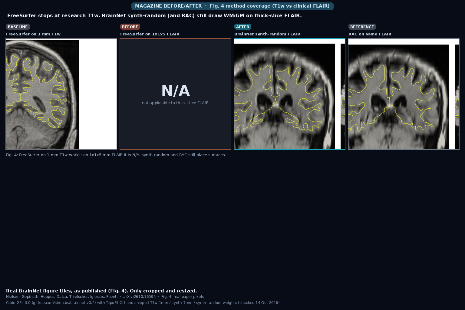
Four coronal panels from Fig. 4: FreeSurfer WM/GM on 1 mm T1w, FreeSurfer N/A on 1x1x5 mm FLAIR, BrainNet synth-random surfaces on the same FLAIR, and RAC on that FLAIR (Nielsen et al., arXiv:2610.16595)

BrainNet: Cortical Surfaces from Any Brain MRI

BrainNet, by Nielsen, Gopinath, Hoopes, Dalca, Thielscher, Iglesias and Puonti (arXiv:2610.16595), reconstructs WM and GM cortical surfaces with vertex-wise uncertainty and fsaverage spherical registration from heterogeneous brain MRI, including thick-slice clinical FLAIR. Inference is about 0.6 s on GPU. Code is GPL-3.0 on GitHub with three TopoFit weight variants.

Read More »
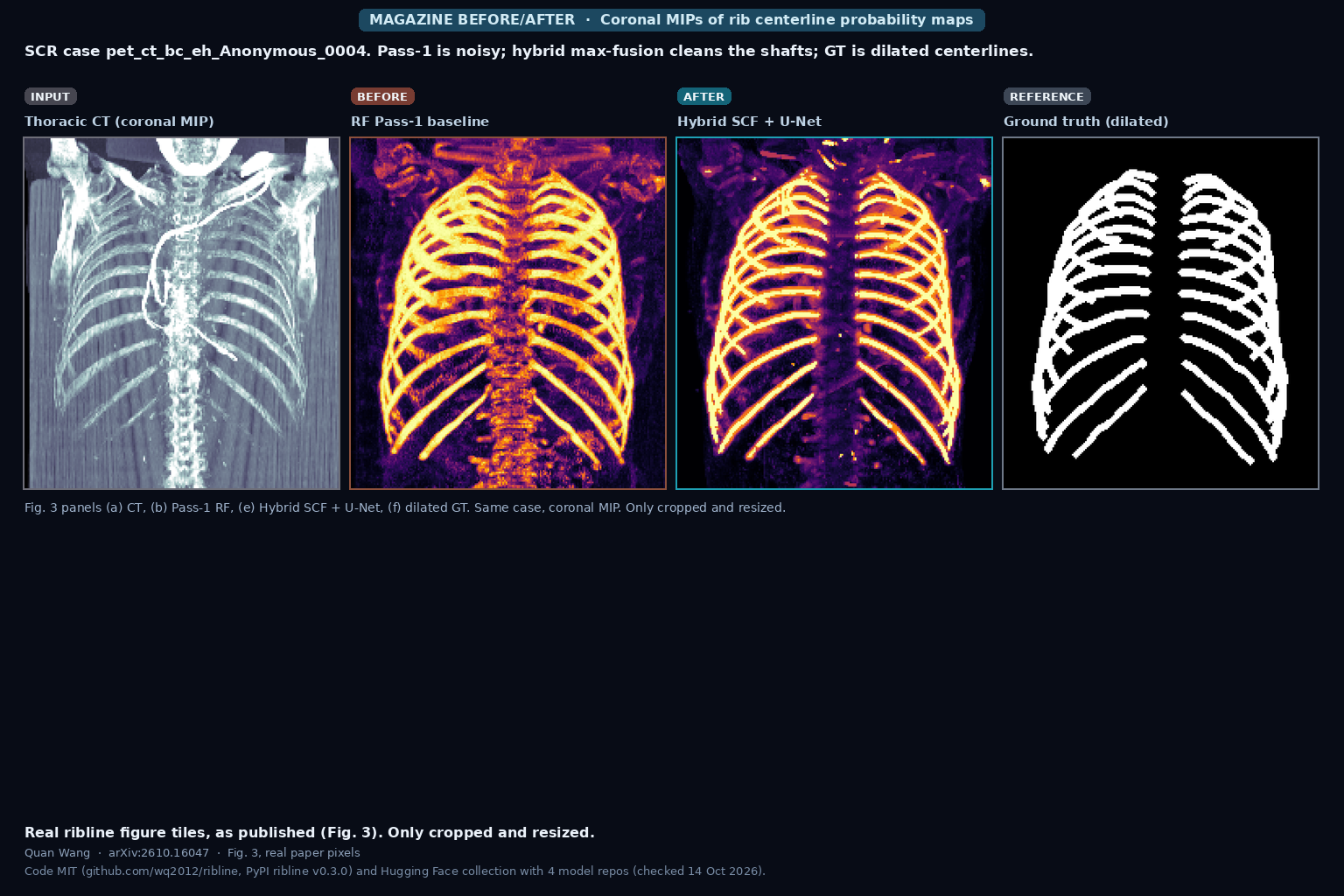
Coronal MIPs of SCR case pet_ct_bc_eh_Anonymous_0004 showing thoracic CT, Pass-1 random-forest probability map, hybrid SCF plus U-Net fusion, and dilated ground-truth rib centerlines (Quan Wang, arXiv:2610.16047, Fig. 3 panels a, b, e, f)

ribline: Numbered 3D Rib Centerlines from Thoracic CT

ribline, by Quan Wang (arXiv:2610.16047), traces and numbers all 24 rib centerlines (R01 to R12, L01 to L12) in thoracic CT with a hybrid Semantic Context Forest plus 3D U-Net detector and bilateral Viterbi matching. On the SCR 2 mm benchmark it reports LCD 2.05 mm and 97.2% labeling accuracy on 12 evaluation scans, with honest caveats on Stage-3 tuning and a 5-fold CV at 3.64 mm / 91.8%. Code is MIT on GitHub and PyPI; weights are on Hugging Face.

Read More »
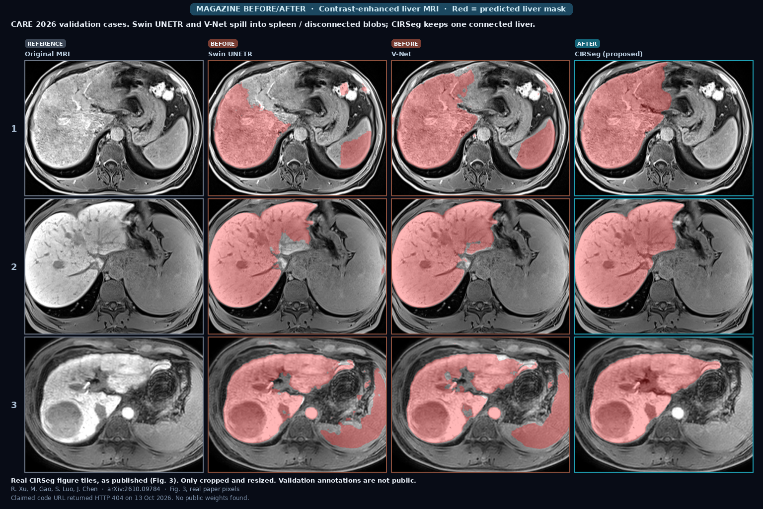
Three CARE 2026 contrast-enhanced liver MRI validation cases with red liver overlays from Swin UNETR and V-Net showing spleen spill and disconnected blobs, beside clean CIRSeg masks and the original images (Xu, Gao, Luo, Chen, arXiv:2610.09784, Fig. 3)

CIRSeg: Liver MRI Masks That Survive Scanner Shift

CIRSeg (Xu, Gao, Luo, Chen) is a cascaded nnU-Netv2 liver segmenter for CARE 2026 contrast-enhanced MRI. It trains with 3D CutMix and Nyul or histogram intensity transfer, refines a coarse prior at full resolution, can adapt at test time without source data, and drops disconnected false positives with LCC filtering. Official CARE eval: Dice 0.9713, HD95 18.31 mm; OOD test 0.9797 / 11.36. Claimed GitHub URL returned HTTP 404 on 13 October 2026. arXiv:2610.09784.

Read More »
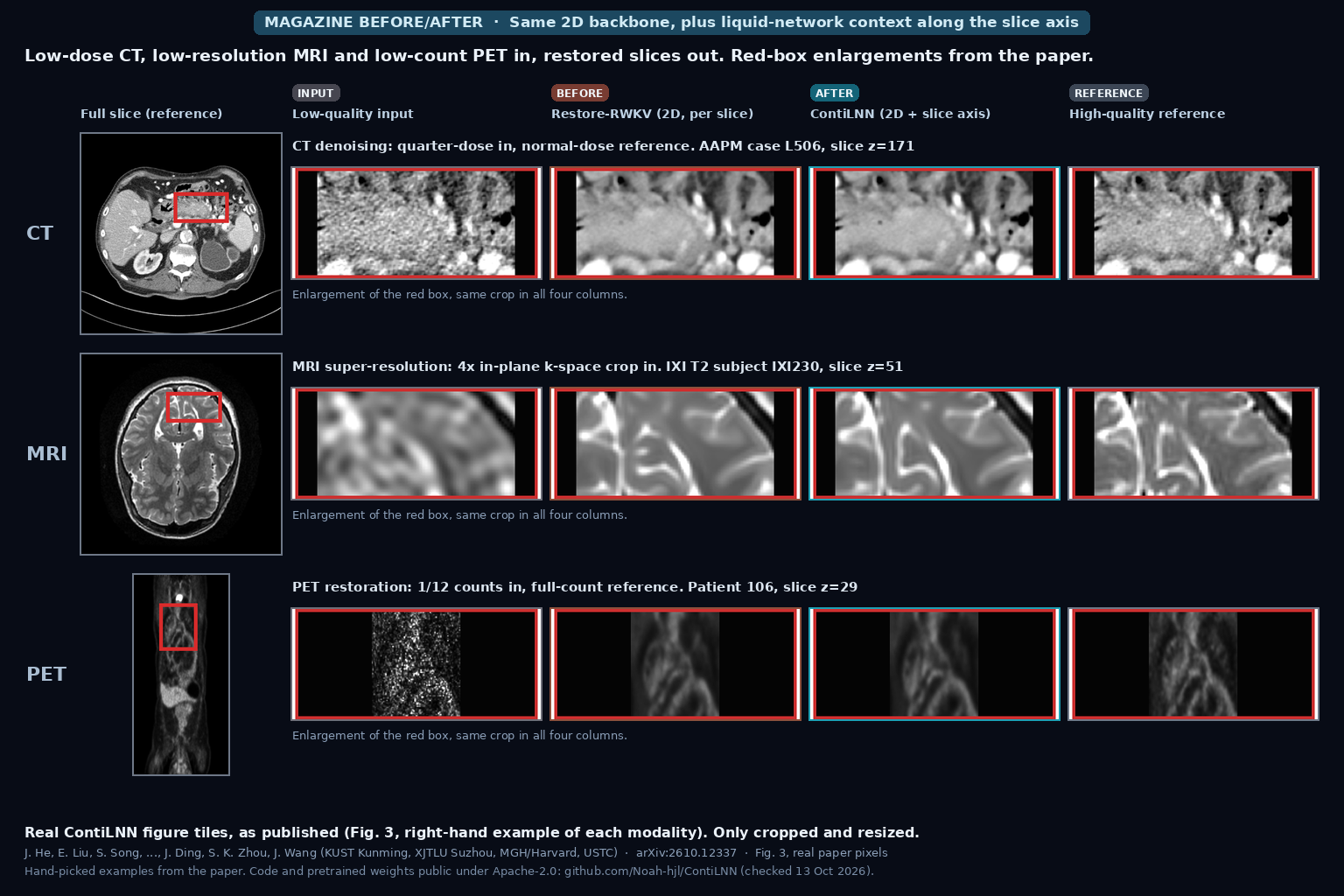
Red-box enlargements of a quarter-dose abdominal CT, a 4x k-space-cropped T2 brain MRI and a 1/12-count PET slice, each shown as low-quality input, Restore-RWKV output, ContiLNN output and high-quality reference, beside the full reference slice (He, Liu, Zhou, Wang et al., arXiv:2610.12337, Fig. 3)

Low-Dose CT, Low-Res MRI or Low-Count PET Slices In, Restored Series Out That Uses Neighboring Slices and Their Gaps, Still Blind to Real DICOM Spacing (ContiLNN)

ContiLNN, from Kunming University of Science and Technology, XJTLU, MGH/Harvard and USTC, adds bidirectional liquid neural network modules along the slice axis of a 2D restoration network, so each restored slice draws on its neighbors and on the index gap to them. Over per-slice Restore-RWKV, mean PSNR rises 1.02 dB for MRI super-resolution and 1.25 dB for low-count PET, the CT gain is small and from one patient, and gaps are counted in slice indices rather than millimeters. Code and weights are public (Apache-2.0). He, Liu, Ding, Zhou, Wang et al., arXiv:2610.12337.

Read More »

We build custom medical imaging platforms — advanced DICOM viewers, AI segmentation, and the clinical systems around them.

Get in Touch

Copyright © 2026 PYCAD. All Rights Reserved.