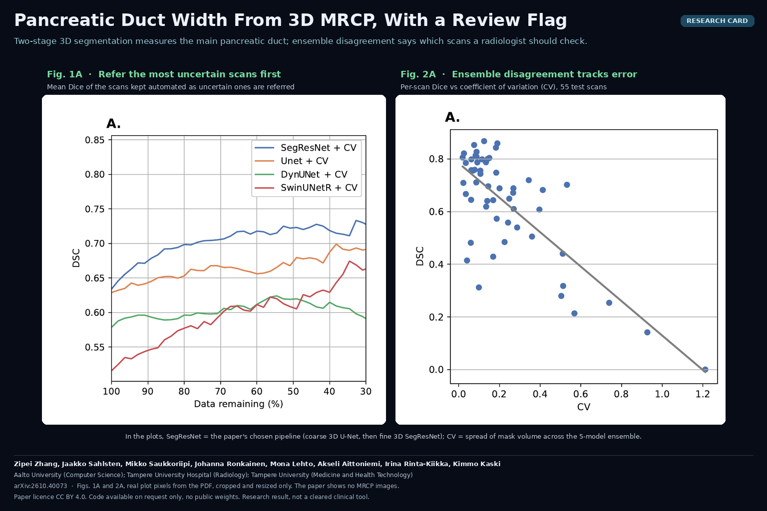
Pancreatic Duct Width From 3D MRCP, With a Review Flag
A two-stage 3D model segments the main pancreatic duct on MRCP, and ensemble disagreement marks the scans a radiologist should check.

A two-stage 3D model segments the main pancreatic duct on MRCP, and ensemble disagreement marks the scans a radiologist should check.
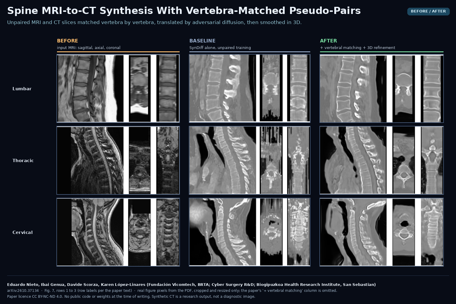
Unpaired MRI and CT slices are matched vertebra by vertebra, translated with adversarial diffusion, then cleaned up in 3D.
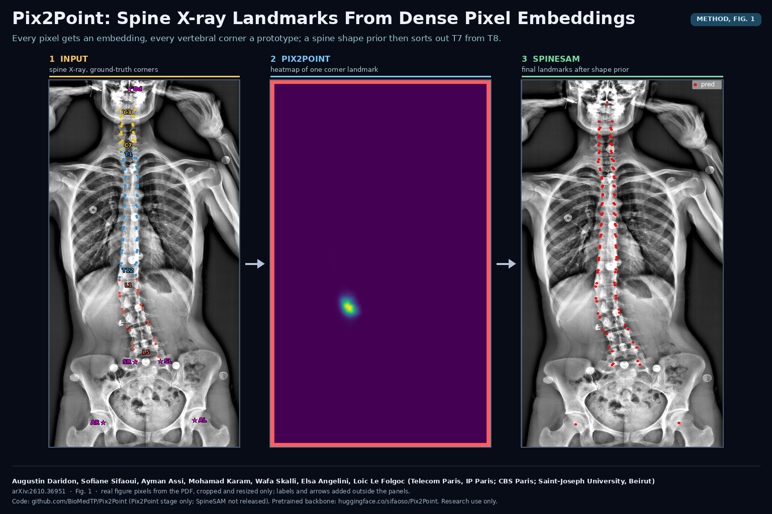
Every pixel gets an embedding and every vertebral corner a prototype; a spine shape prior then decides which corner is which.
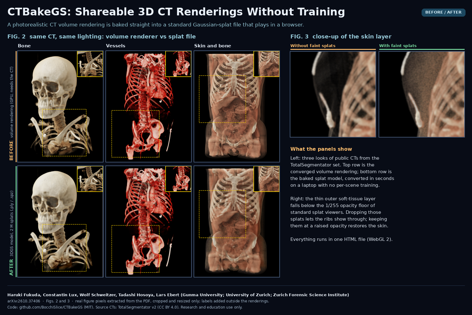
A browser tool bakes lit CT volume renderings into standard Gaussian-splat files in seconds, with no training and MIT code.
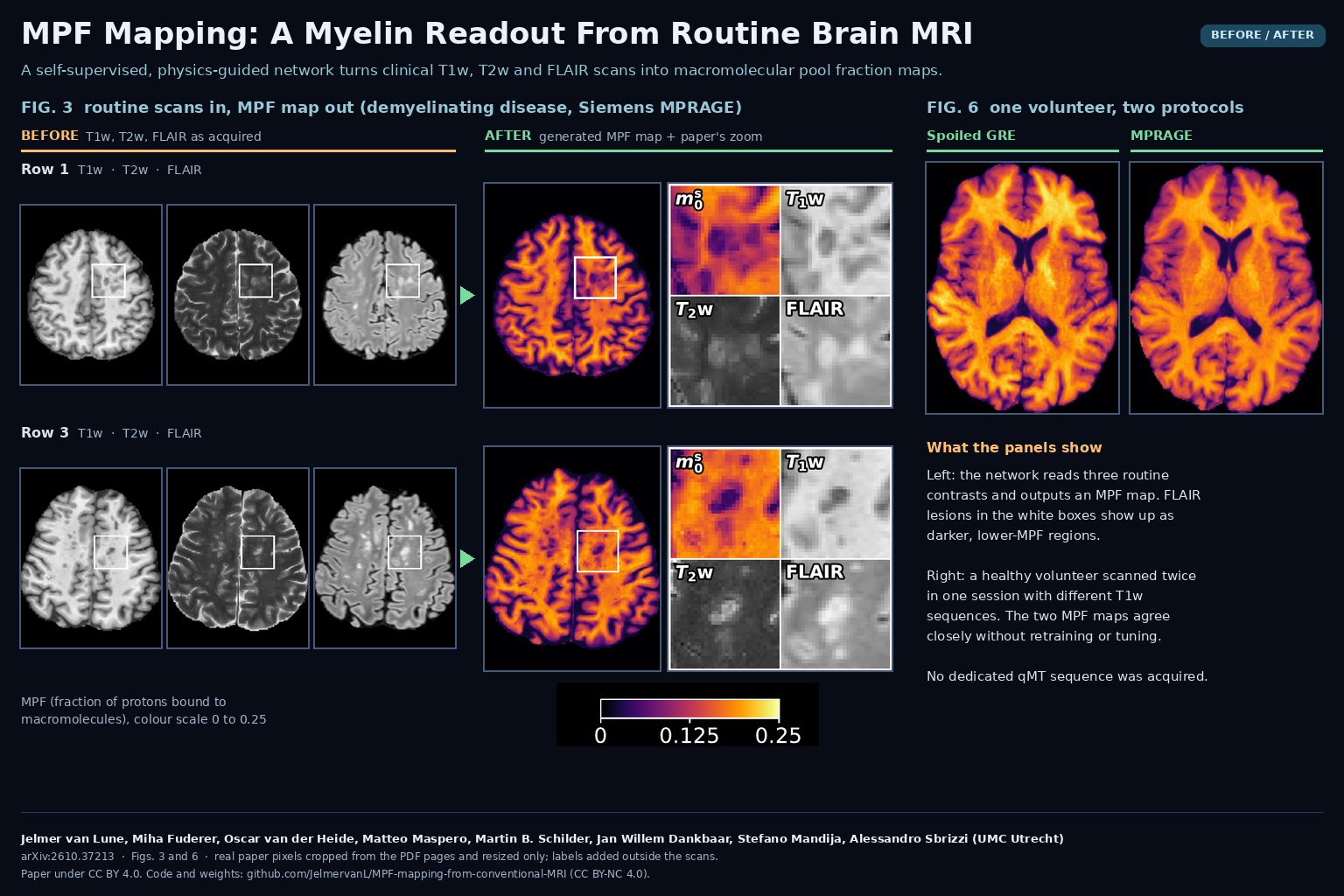
A self-supervised network turns T1w, T2w and FLAIR scans into macromolecular pool fraction maps, with public code and weights.
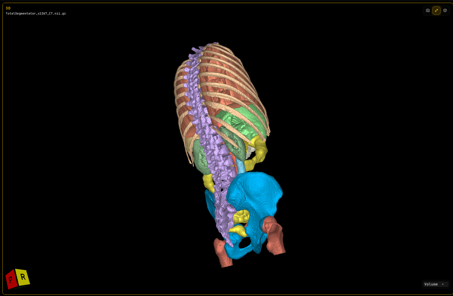
Send clients the CT or MRI scan, label maps and 3D meshes in one browser link that opens on desktop or phone, with nothing to install. Try the live demo.