Generic selectors
Exact matches only
Search in title
Search in content
Post Type Selectors
PYCAD BLOG

Insights on Medical Imaging, AI & DICOM

Guides, project breakdowns, and lessons learned from building medical imaging platforms — written by the PYCAD team.

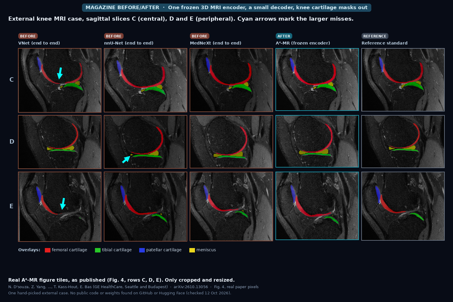
Sagittal knee MRI slices with femoral, tibial, patellar cartilage and meniscus overlays from VNet, nnU-Net, MedNeXt, A4-MR and the reference standard, cyan arrows on larger misses (D'souza, Bas et al., arXiv:2610.13056, Fig. 4)

Whole 3D MRI Series In, Prompt-Free Organ Boxes and Report-Searchable Anatomy Out From One Frozen Encoder, Still Weak on Organs Smaller Than Its 8-Voxel Patches (A4-MR)

A4-MR, a 3D MRI vision-language model from GE HealthCare trained on about 50,000 studies (491,000 series), finds 125 anatomy classes as 3D boxes without prompts, ties each box to report text, and keeps healthy and abnormal organs apart in its embedding. Zero-shot box IoU on AMOS MR is 66.3 against 65.5 for MedSAM2 given a ground-truth box, small organs stay hard, and no code or weights are public. D’souza, Bas et al., arXiv:2610.13056.
Read More »
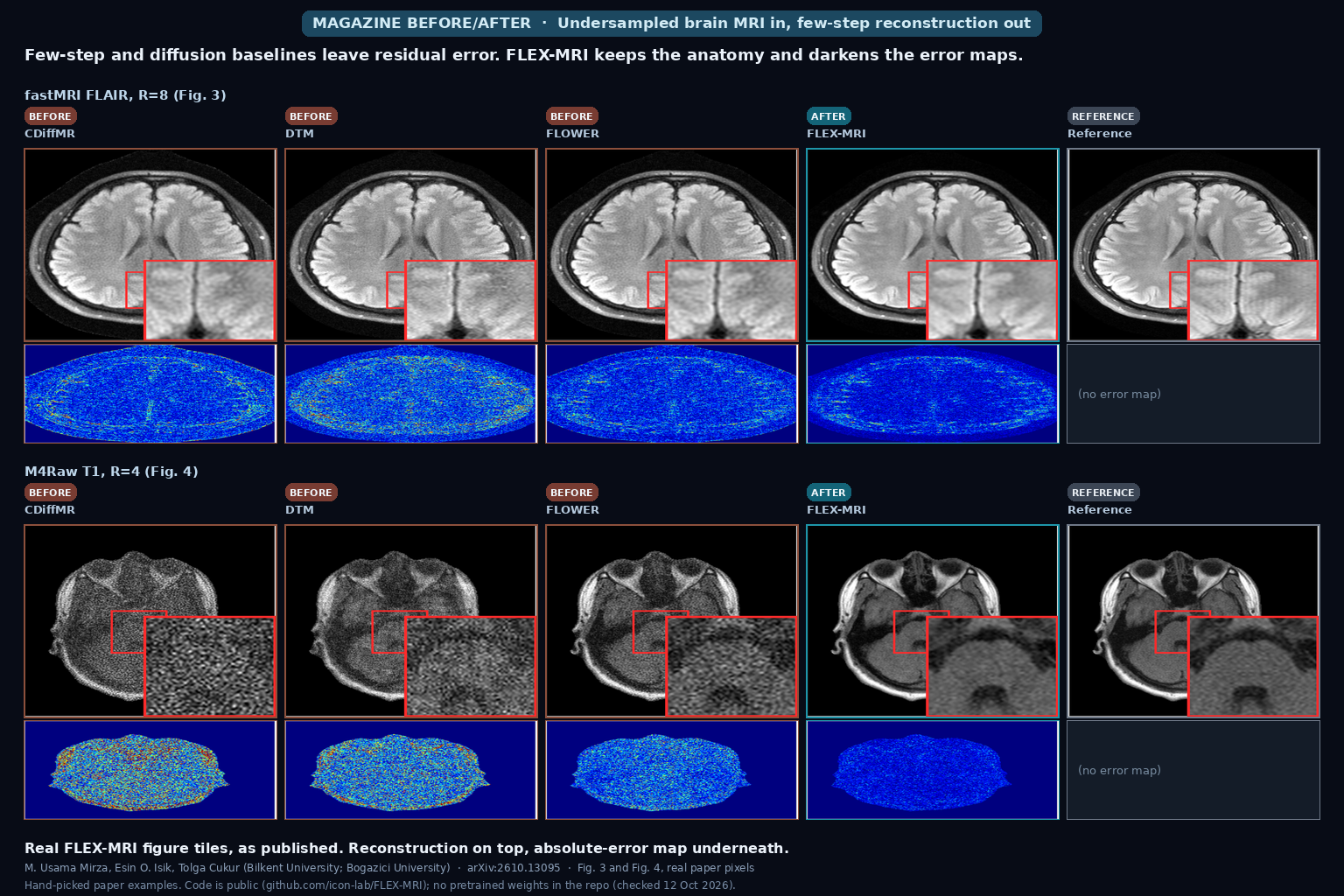
Brain MRI reconstructions at high acceleration: three few-step or diffusion baselines, FLEX-MRI, and the fully sampled reference, each with absolute-error maps (Mirza, Isik, Cukur, arXiv:2610.13095, Fig. 3 and Fig. 4)

Undersampled Brain MRI In, Few-Step Reconstruction Out That Explores Alternate k-Space Paths Without Hundreds of Diffusion Steps

FLEX-MRI takes undersampled multi-coil brain MRI and recovers the image in about ten finite transitions with stochastic k-space exploration and data consistency, without a 1000-step diffusion sampler. On fastMRI at R=8 it reaches 35.8 dB PSNR and 92.7% SSIM, runs in 0.349 s with 1.05 GB on an RTX 4090, and the GitHub repo has training and eval code but no pretrained weights. Mirza, Isik, Cukur, arXiv:2610.13095.

Read More »
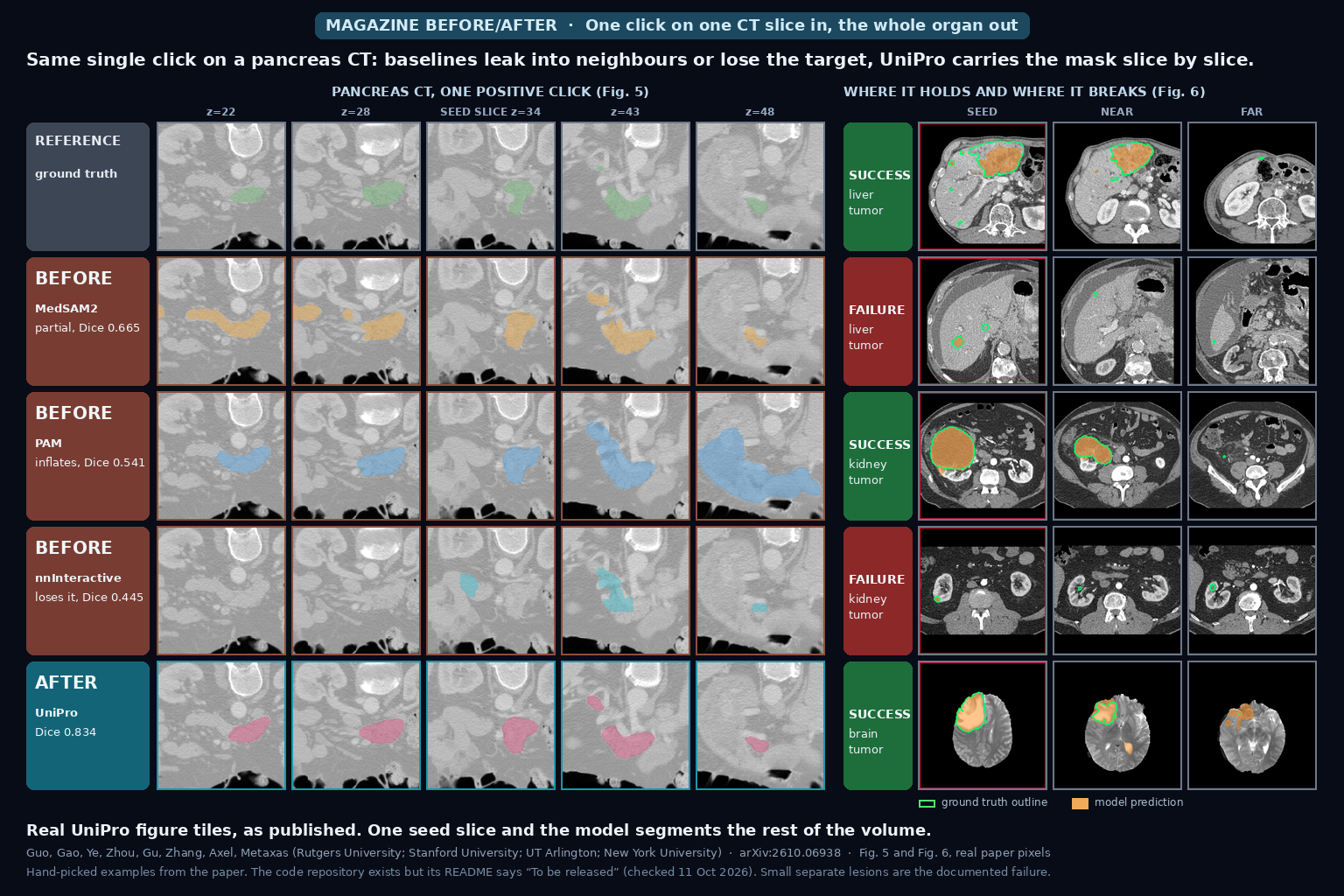
Pancreas CT slices after one click: ground truth, three baseline propagation models and UniPro at five slice positions, with liver, kidney and brain tumor success and failure examples on the right (Guo et al., arXiv:2610.06938, Fig. 5 and Fig. 6)

One Click on One CT Slice In, a Full 3D Organ Mask Out That Is Carried Slice by Slice From the Neighbours and Still Takes Class Prompts, Reference Images and Corrections in the Same Model

Click once on one slice of a CT or MR volume and UniPro carries the mask through the other slices, using the seed slice and the latest predictions as references. The same 2D model also takes a class name, a reference example or corrections. It is best of the compared models on 7 of 8 single-click tests, trails 3D nnU-Net in plain semantic mode, needs 28 seconds per abdominal volume, and the repository README still says “To be released”. Guo et al., arXiv:2610.06938.

Read More »
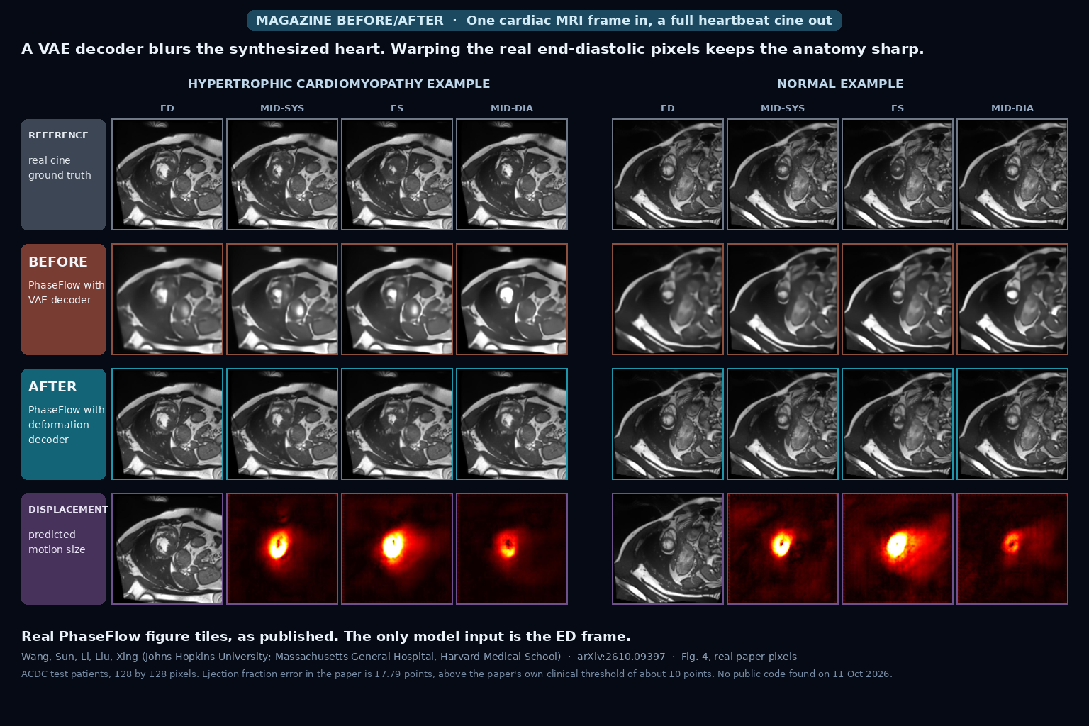
Cardiac MRI cine frames at end-diastole, mid-systole, end-systole and mid-diastole for a hypertrophic cardiomyopathy and a normal example: real cine on top, a blurry VAE-decoder result in the middle, a sharp deformation-decoder result below, and predicted displacement heat maps at the bottom (Wang et al., arXiv:2610.09397, Fig. 4)

One End-Diastole Cardiac MRI Frame In, a Full 30-Frame Beating-Heart Cine Out Without ECG Gating by Warping the Real Pixels With a Phase-Conditioned Flow Model

Give PhaseFlow one end-diastolic cardiac MRI frame and it generates a 30-frame cine of the heartbeat with no ECG gating. It predicts a motion field and warps the real pixels, which keeps the image sharp, and it takes its timing from a mean phase curve for the patient’s pathology group. Tested on ACDC with 30 test patients and one training run, the ejection fraction error is 17.79 points, above the paper’s own clinical threshold of about 10, and I found no public code. Wang et al., arXiv:2610.09397.

Read More »
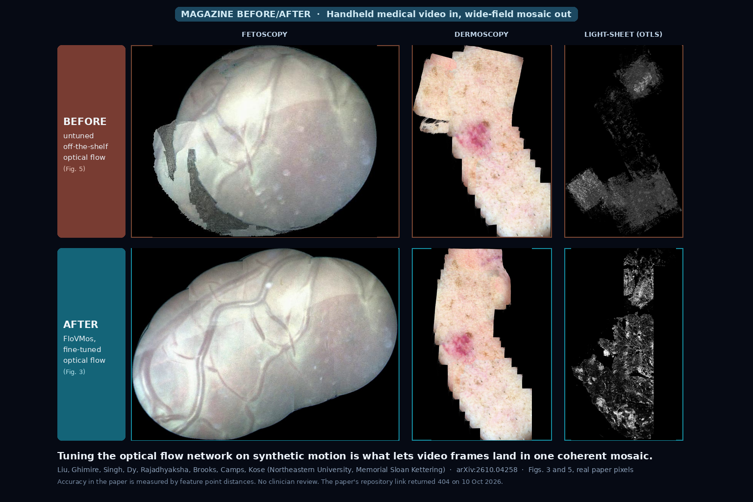
Before and after mosaics of fetoscopy, dermoscopy and light-sheet microscopy video: the top row, stitched with an untuned optical flow model, shows grey patches and broken pieces; the bottom row, from the fine-tuned FloVMos model, is continuous (Liu et al., arXiv:2610.04258, Figs. 3 and 5)

Handheld Endoscope or Microscope Video In, One Wide-Field Tissue Mosaic Out at About Ten Frames per Second From a Modality-Tuned Optical Flow Model

Feed FloVMos video from a handheld endoscope, dermoscope or confocal microscope and it stitches the frames into one growing wide-field mosaic, at about ten frames per second on 1 megapixel frames in the authors’ research code. The trick is fine-tuning an optical flow network per device on synthetic motion, so no hand-labelled flow is needed. Several real test sets are one or two videos, accuracy is a feature distance check, a GPU is required, and the repository linked in the paper returned a 404 when I checked. Liu et al., arXiv:2610.04258.

Read More »
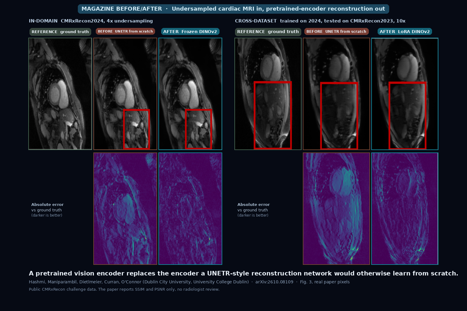
Two panels of cardiac MRI slices with absolute error maps below, each showing the ground truth, a UNETR trained from scratch, and a reconstruction from a pretrained DINOv2 encoder, at 4x acceleration in-domain and 10x acceleration across datasets (Hashmi et al., arXiv:2610.08109, Fig. 3)

Undersampled Cardiac MRI In, Sharper Reconstruction Out From a Frozen Pretrained Vision Encoder With About Half the Trainable Weights

Hand an undersampled cardiac MRI slice to a UNETR-style network whose encoder is a frozen pretrained CLIP, BiomedCLIP or DINOv2 model, and the reconstruction beats the same network trained from scratch, with about 49 percent fewer trainable parameters. The gap holds with 5 percent of the training data and across two challenge datasets. It is a 2D study on public CMRxRecon data scored with SSIM and PSNR only, with no reader study, and the linked GitHub repository held only a README when I checked. Hashmi et al., arXiv:2610.08109.

Read More »

We build custom medical imaging platforms — advanced DICOM viewers, AI segmentation, and the clinical systems around them.

Get in Touch

Copyright © 2026 PYCAD. All Rights Reserved.