
Brain T1 Plus Tumor Mask In, Slice-Consistent Healthy Tissue Fill Out for Registration and Parcellation
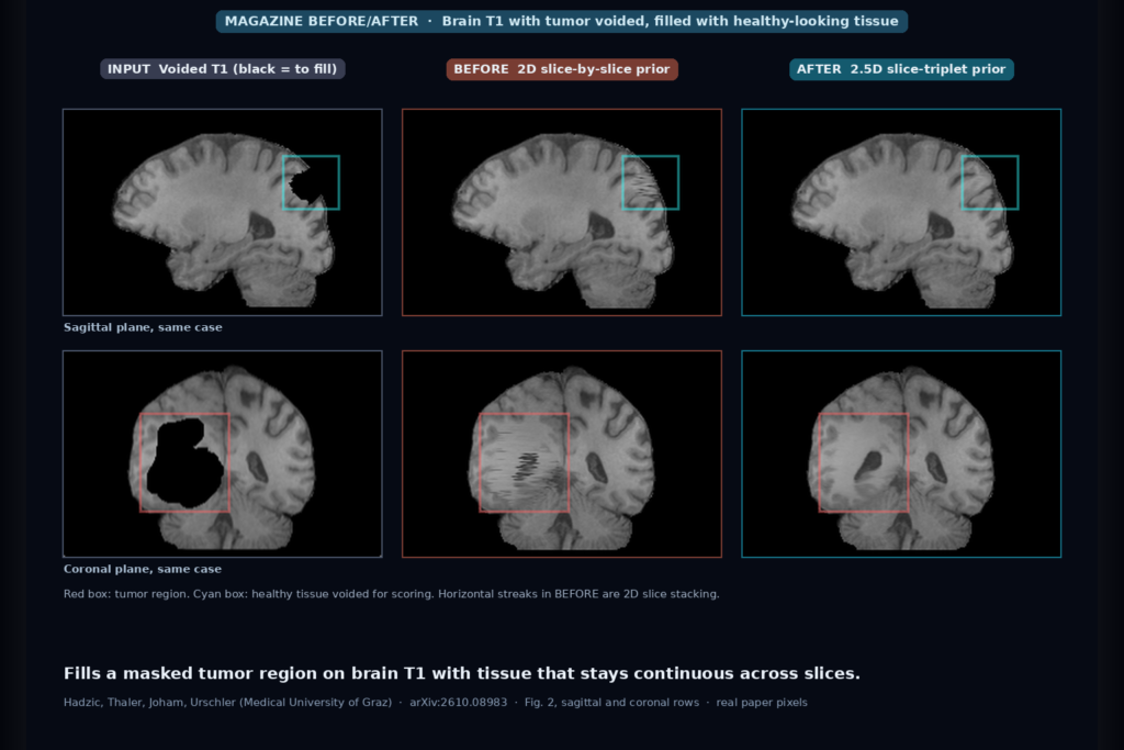
Give it a brain T1 and a mask over the tumor, and it paints in healthy-looking tissue that stays continuous across slices, so atlas registration and parcellation can run on a filled copy. Code and checkpoint public. Hadzic et al., arXiv:2610.08983.
Prostate MRI Near Rectal Gas or Metal In, Undistorted High-Resolution DWI and ADC Out
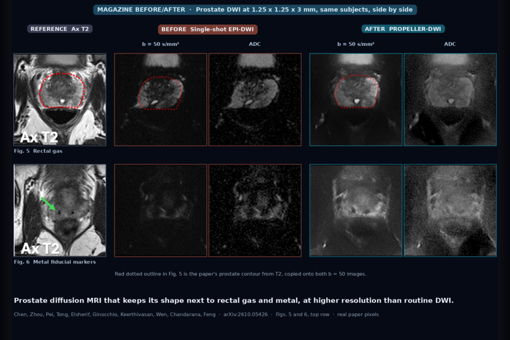
A PROPELLER diffusion sequence with self-supervised reconstruction keeps the prostate’s shape on DWI and ADC next to rectal gas and fiducial markers, at 1.25 mm in-plane. Siemens prototype, no public code. Chen et al., arXiv:2610.05426.
3D T1 Brain MRI In, Closest Real Scans Out, Searchable by Whole Brain or Any of 103 Regions
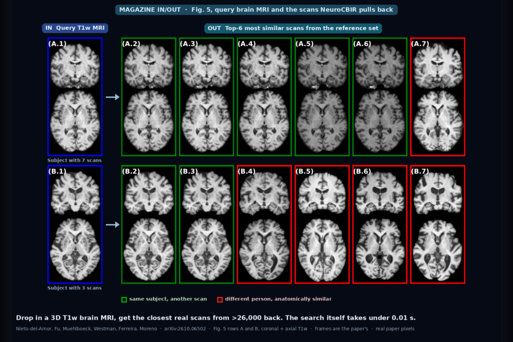
Drop in a 3D T1w brain MRI and get back the most anatomically similar real scans from a reference set of more than 26,000, by whole brain or by region. Code, weights, and embeddings public. Nieto-del-Amor et al., arXiv:2610.06502.
On-Board Radiotherapy CBCT In, Cleaner CT-Like Slices Out, Trained Without Paired Scans
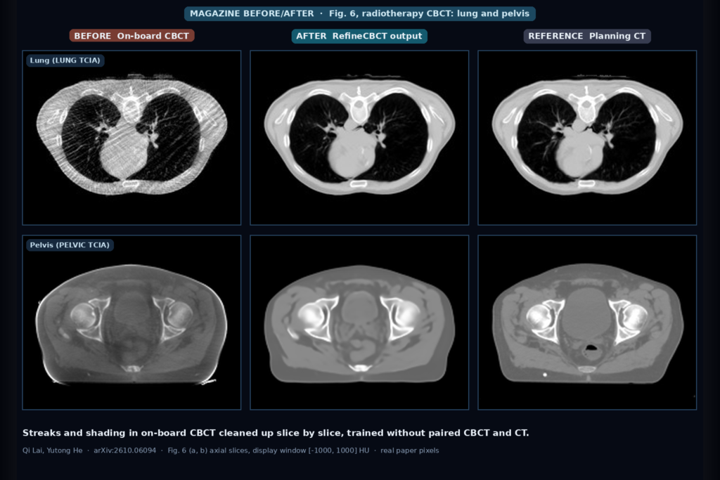
Feed it an on-board radiotherapy CBCT slice and get back one with fewer streaks and CT numbers closer to the planning CT. RefineCBCT learns without paired CBCT and CT. Code public. Lai and He, arXiv:2610.06094.
Abdominal CT Report Sentence In, 3D Finding Mask Out, Learned From the Arrows and Measurement Lines Already in PACS
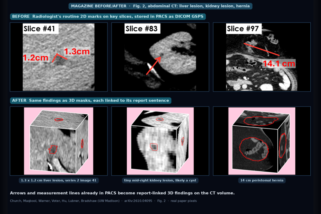
Give it one report sentence and an abdominal CT; get a 3D mask of that finding back. LocusCT learns from 105K routine PACS arrows and measurement lines matched to report text. Church et al., arXiv:2610.04095.
Segmented CT or MR In, Real-Time Cinematic 3D Out That Recolors and Hides Tissue Without Refitting
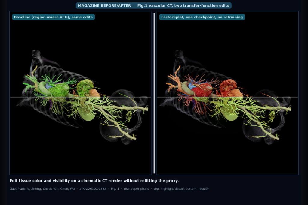
Hang a segmented CT or MR in; get a real-time cinematic 3D proxy out whose tissue colors, opacity, and visibility you can change without refitting. Gao et al., arXiv:2610.02382.
Reconstructed Implant CT In, Streak-Reduced Tissue Out Without Raw Projections or Training
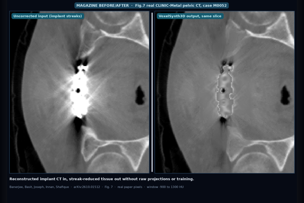
Hang a reconstructed implant CT in; get a streak-reduced derived series out with no raw projections, vendor kernel, or trained weights. Banerjee et al., arXiv:2610.01512.
Multimodal MRI In; ET, RC, TC, and WT Overlays Out With RC-Aware Pre vs Post Routing
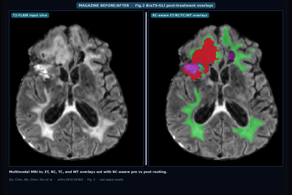
Hang T1/T1Gd/T2/T2-FLAIR glioma MRI in; get ET, RC, TC, and WT overlays out with treatment-status routing. BraTS-GLI 2025 2nd place (ECNU NYU). arXiv:2610.01060.
CTA, MRA, and Routine Brain MRI In; Aneurysm Labels, Crosshair Localizers, and 13-Vessel Overlays Out
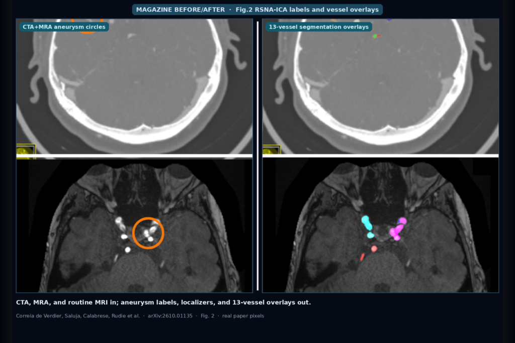
Hang CTA, MRA, and routine brain MRI from RSNA-ICA: aneurysm presence and 13-location labels, crosshair localizers, plus 178-series vessel NIfTI overlays. 7202 series, 21 sites. arXiv:2610.01135.
SWAN Stack In, Synthetic-Trained Microbleed Overlays Out When Real CMB Labels Are Scarce
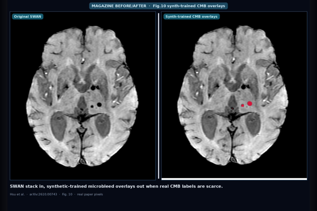
Hang a SWAN stack into a neuro MRI workflow: synthetic soft-ellipsoid CMBs on real negative hosts train a MONAI 3D U-Net that keeps ~84% Dice / ~88% lesion sensitivity vs real-trained when CMB labels are scarce. Hsu et al., arXiv:2610.00743.