Skip to content
  • Home
  • Services
    • All Services
    • Custom DICOM Viewer
    • Case Studies
  • Products
    • All Products
    • PYCAD DICOM Viewer
    • PYCAD Dental Viewer
    • PYCAD SSR Engine
  • Case Studies
  • Blog
  • About us
  • Home
  • Services
    • All Services
    • Custom DICOM Viewer
    • Case Studies
  • Products
    • All Products
    • PYCAD DICOM Viewer
    • PYCAD Dental Viewer
    • PYCAD SSR Engine
  • Case Studies
  • Blog
  • About us
Get in Touch

Category: machine learning

SIM: Zero-Shot MRI Reconstruction Learned From Simulated Data
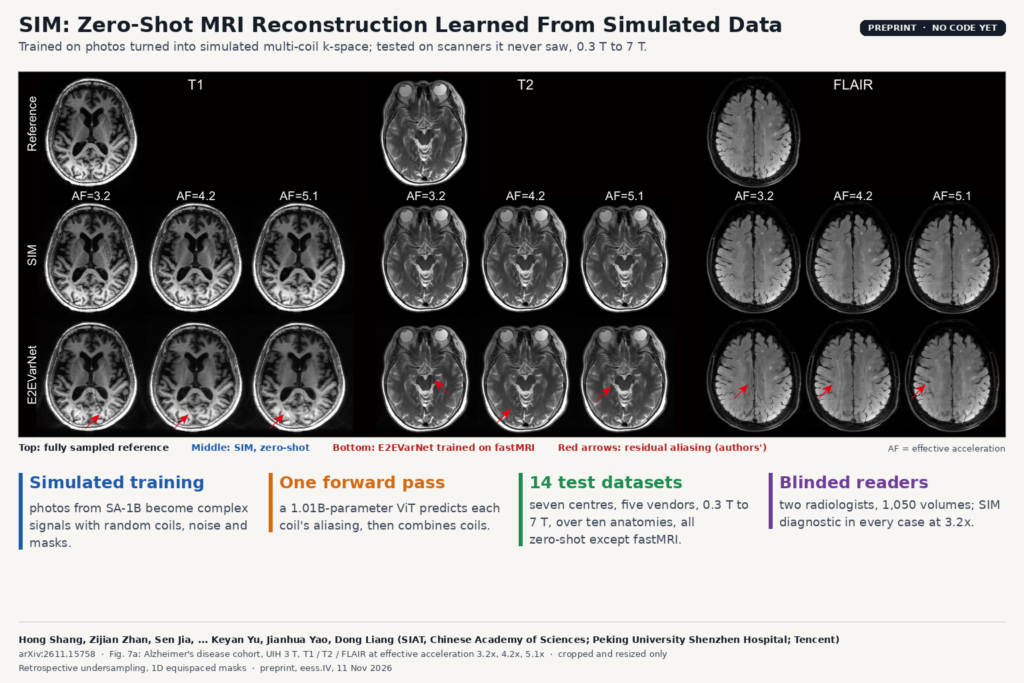
Fig. 7a of arXiv:2611.15758 by Shang, Zhan, Jia and co-authors (SIAT Chinese Academy of Sciences, Peking University Shenzhen Hospital, Tencent): T1, T2 and FLAIR brain MRI from an Alzheimer's disease cohort, fully sampled reference above zero-shot SIM and E2EVarNet reconstructions at three accelerations, red arrows on residual aliasing.

A one-billion-parameter transformer trained on simulated k-space from photos reconstructs MRI from scanners it never saw, 0.3 T to 7 T.

X-Strata: Separating Aneurysm and Vessel Layers in 2D Angiography
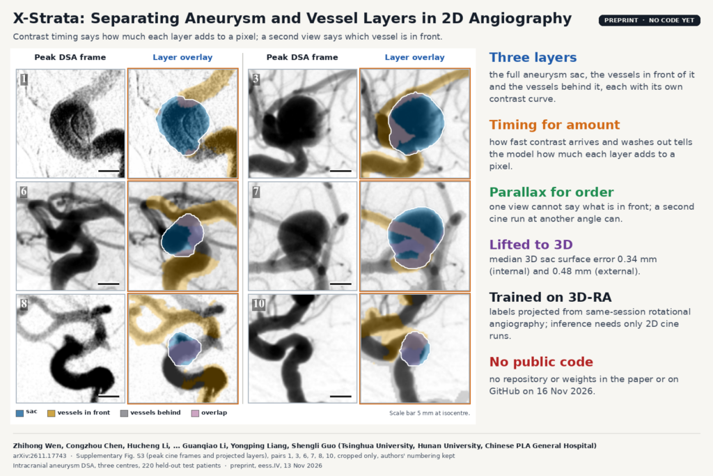
Supplementary Fig. S3 of arXiv:2611.17743 by Wen, Chen, Li and co-authors (Tsinghua University, Hunan University, Chinese PLA General Hospital): six pairs of peak cine DSA frames of intracranial aneurysms next to layer overlays with the sac in blue, vessels in front in gold, vessels behind in gray and overlap in pink.

From two routine DSA cine runs, X-Strata recovers the full aneurysm sac, sorts crossing vessels in front or behind, and lifts both to 3D.

AgentCT: A CT Agent That Picks the Right Restoration Model
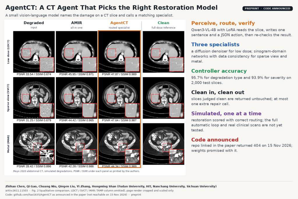
Fig. 2 of arXiv:2611.11503 by Chen, Gao, Niu, Liu, Zhang and Shan (Fudan University and co-authors): abdominal CT slices with simulated low-dose noise, sparse-view streaks and metal artifacts, comparing the degraded input, the all-in-one AMIR model, the AgentCT routed specialist and the clean reference, with zoomed regions and PSNR/SSIM.

A small vision-language model reads a CT slice, names the damage and sends it to a low-dose, sparse-view or metal specialist.

DuSS-PET: Low-Dose PET Reconstruction Without Full-Dose Pairs
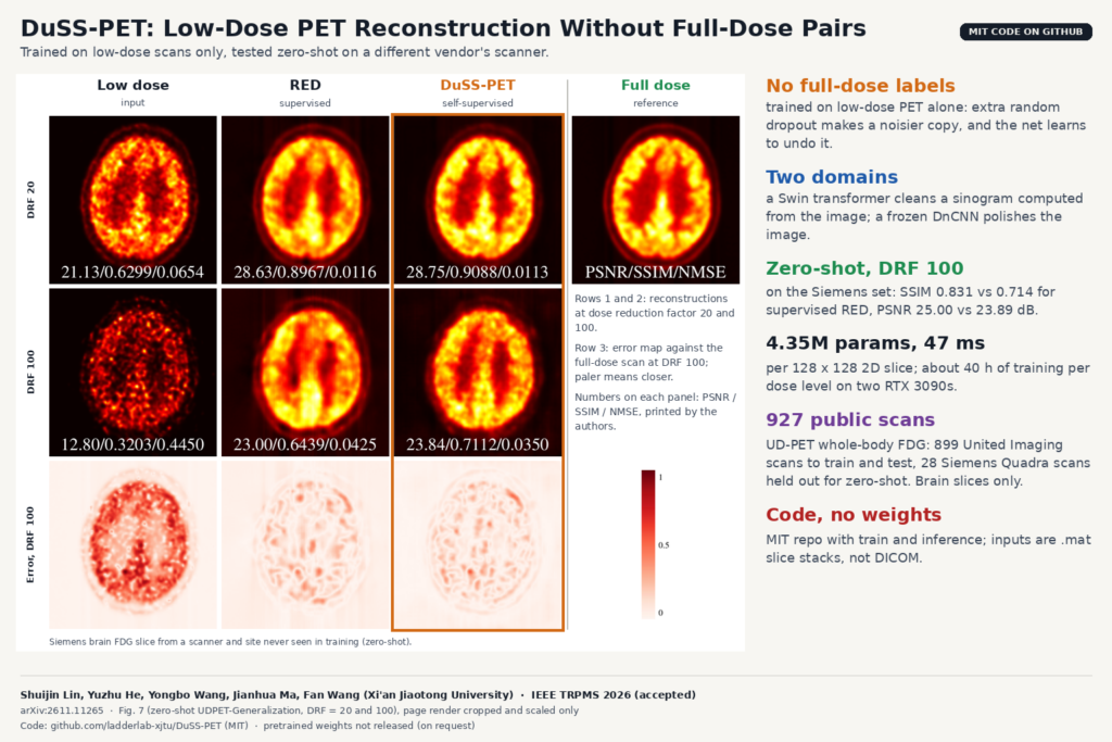
Fig. 7 of arXiv:2611.11265 by Lin, He, Wang, Ma and Wang (Xi'an Jiaotong University): zero-shot brain FDG PET reconstructions on a Siemens scanner at dose reduction factors 20 and 100, comparing the low-dose input, the supervised RED model and the self-supervised DuSS-PET with the full-dose reference, plus error maps.

A self-supervised model learns to clean low-dose PET from low-dose scans alone and holds up on another vendor’s scanner.

GenStrain: DENSE-Quality Myocardial Strain From Routine Cine MRI
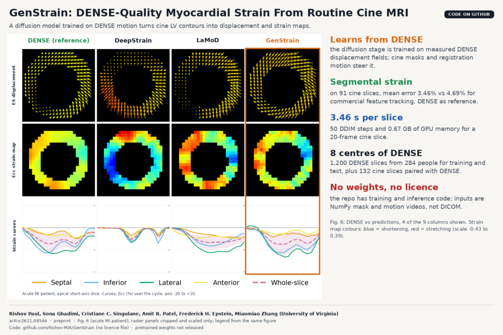
Fig. 6 of arXiv:2611.08544 by Paul, Ghadimi, Singulane, Patel, Epstein and Zhang (University of Virginia): end-systolic displacement fields, circumferential strain maps and segmental strain curves for one acute myocardial infarction patient, comparing the DENSE reference with DeepStrain, LaMoD and GenStrain.

A diffusion model trained on DENSE motion predicts myocardial displacement and strain maps from standard cine cardiac MRI.

OneStepBrainMRI: Forecasting Brain Atrophy From One Baseline MRI
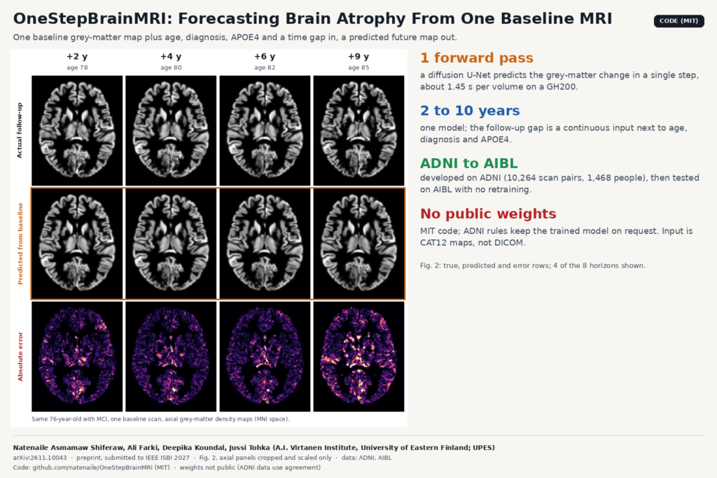
Fig. 2 of arXiv:2611.10043 by Shiferaw, Farki, Koundal and Tohka (University of Eastern Finland): axial grey-matter density maps of one ADNI participant with MCI, showing the measured follow-up, the one-step diffusion prediction from a single baseline scan, and the absolute error at +2, +4, +6 and +9 years.

A one-step diffusion model predicts grey-matter maps 2 to 10 years ahead from one baseline MRI, age, diagnosis and APOE4.

moco-dl: Fast Slice-Wise Motion Correction for Spinal Cord fMRI
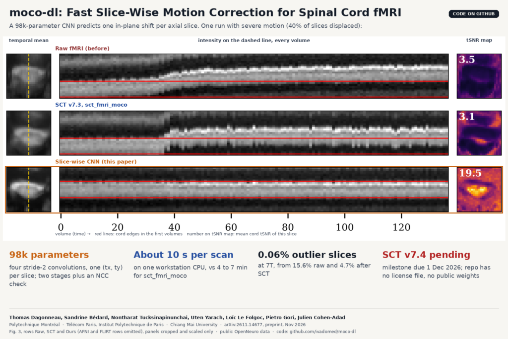
Fig. 3 of arXiv:2611.14677 by Dagonneau, Bedard et al. (Polytechnique Montreal, Telecom Paris, Chiang Mai University): temporal mean, intensity profile over time and tSNR map of one spinal cord fMRI slice with severe motion, for the raw data, SCT sct_fmri_moco and the proposed slice-wise CNN.

A tiny CNN aligns every axial slice of a spinal cord fMRI run on a CPU in seconds and beats SCT’s current tool at 3T and 7T.

LoopMR: Accelerated MRI Reconstruction With One Looped Block
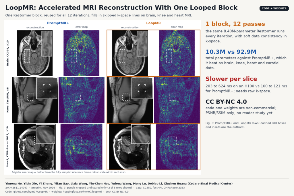
Fig. 3 of arXiv:2611.14947 by He, Xie et al. (Cedars-Sinai Medical Center): PromptMR+ and LoopMR reconstructions of undersampled brain (CC359 x10), knee (fastMRI x8) and heart (CMRxRecon2023 x10) MRI with their absolute error maps and the authors' zoomed regions of interest.

One Restormer block, run 12 times, rebuilds undersampled brain, knee, heart and carotid MRI with a ninth of PromptMR+’s parameters.

CADS: One Open Model Suite for 167 Whole-Body CT Structures
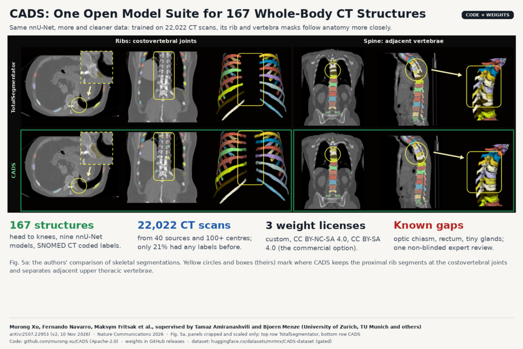
Fig. 5a of arXiv:2507.22953 by Xu et al. (University of Zurich, TU Munich and others): CT slices and 3D renders comparing TotalSegmentator (top) and CADS (bottom) rib and vertebra masks, with the authors' yellow circles at the costovertebral joints and upper thoracic vertebrae.

Trained on 22,022 curated CT scans from 100+ centres, CADS segments head to knees, runs in 3D Slicer and outputs SNOMED CT labels.

MicroBT: Finding and Counting Sub-3 mm Brain Tumors on MRI
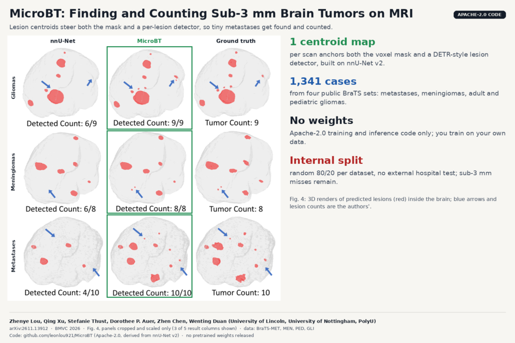
Fig. 4 of arXiv:2611.13912 by Lou, Xu et al. (University of Lincoln, University of Nottingham, PolyU): 3D brain renders with red tumor lesions predicted by nnU-Net and MicroBT next to ground truth, with the authors' lesion counts and blue arrows marking small lesions.

A centroid-guided nnU-Net extension segments brain tumors on MRI and counts each lesion, aimed at the tiny metastases that models miss.

← Previous
Next →