Skip to content
  • Home
  • Services
    • All Services
    • Custom DICOM Viewer
    • Case Studies
  • Products
    • All Products
    • PYCAD DICOM Viewer
    • PYCAD Dental Viewer
    • PYCAD SSR Engine
  • Case Studies
  • Blog
  • About us
  • Home
  • Services
    • All Services
    • Custom DICOM Viewer
    • Case Studies
  • Products
    • All Products
    • PYCAD DICOM Viewer
    • PYCAD Dental Viewer
    • PYCAD SSR Engine
  • Case Studies
  • Blog
  • About us
Get in Touch

Category: Medical Imaging

LAPIS: Chest X-Ray Anonymization Beyond the DICOM Header
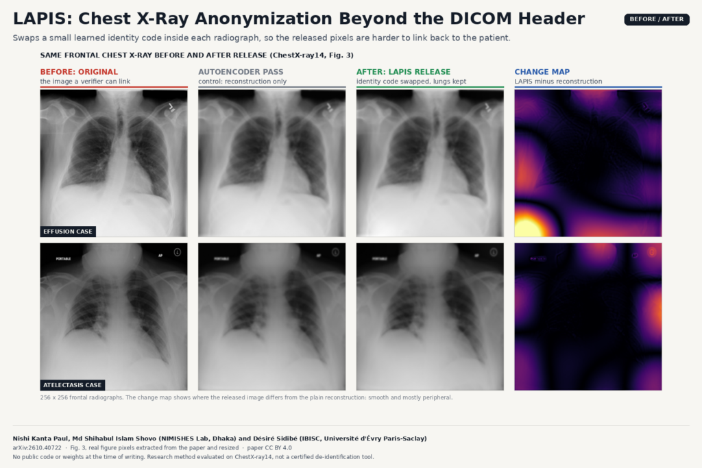
Before and after figure built from Figure 3 of arXiv:2610.40722 by Nishi Kanta Paul, Md Shihabul Islam Shovo and Désiré Sidibé: two frontal chest radiographs (effusion and atelectasis cases) shown as original, autoencoder reconstruction, LAPIS anonymized release and a change map that is smooth and mostly peripheral.

LAPIS swaps a small learned identity code inside each chest X-ray so released images are harder to link back to the patient.

EUReCA: One Network for Sparse-View Linac CBCT
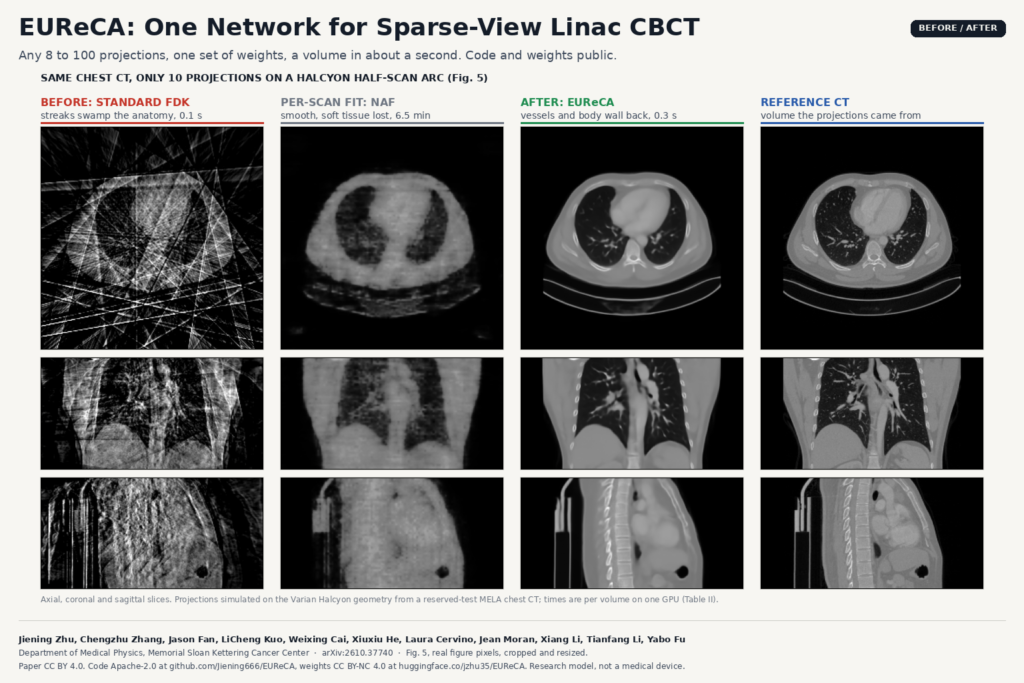
Before and after figure built from Figure 5 of arXiv:2610.37740 by Jiening Zhu, Yabo Fu and colleagues (Memorial Sloan Kettering Cancer Center): axial, coronal and sagittal chest slices reconstructed from 10 projections with standard FDK (streaks), NAF per-scan fitting (smooth, soft tissue lost), EUReCA (vessels and body wall recovered) and the reference CT.

Any 8 to 100 projections on a Halcyon arc, one set of weights, and a CBCT volume in about a second instead of minutes.

A-Fuse: Airway Tree Segmentation That Holds Up on Pediatric CT
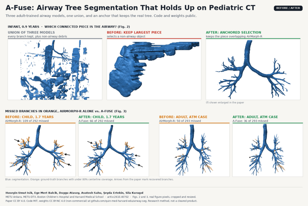
Before and after figure built from Figures 2 and 3 of arXiv:2610.40702 by Huseyin Umut Isik, Sila Kurugol and colleagues (METU and Boston Children's Hospital): for a 0.9-year-old infant, the union of three airway models full of non-airway debris, the largest connected piece that is not the airway, and the anchored selection that keeps the airway tree; below, 3D airway renders for a child and an adult comparing AirMorph-R alone with A-Fuse, with missed ground-truth branches in orange.

Three adult-trained airway models, one union, and an anchor rule that keeps the real tree on child and adult chest CT.

LesionStudio: Tune PET/SPECT Reconstruction With Synthetic Lesions
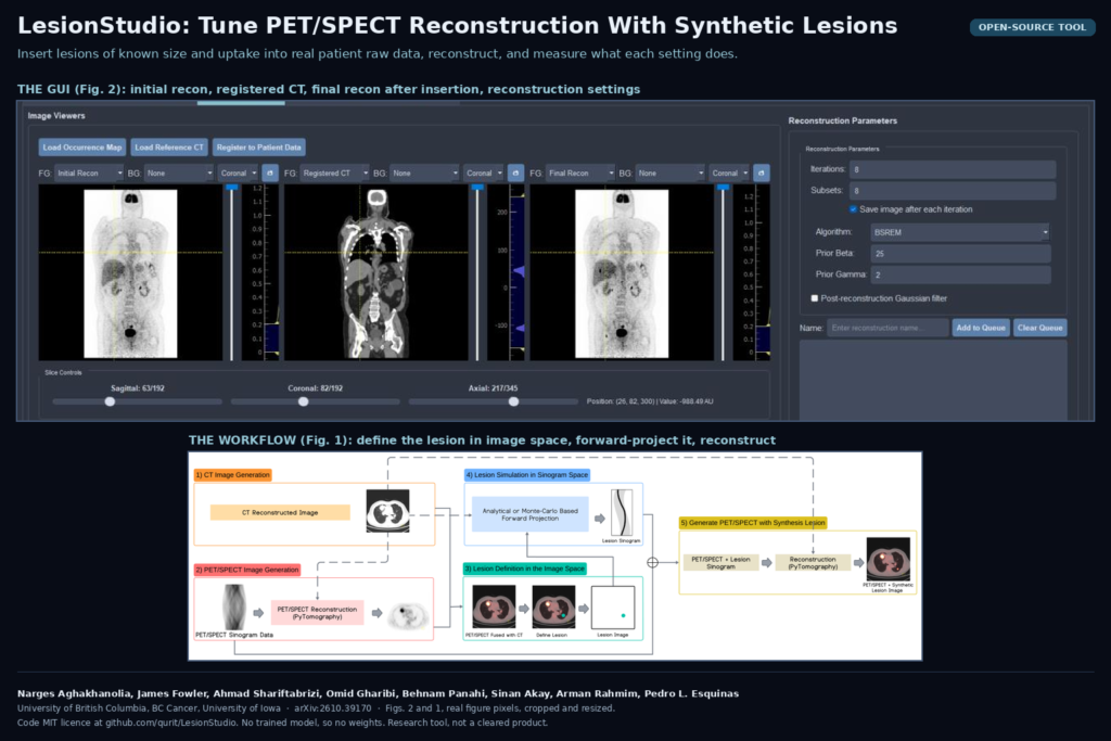
Banner from Figures 2 and 1 of arXiv:2610.39170 by Narges Aghakhanolia, Pedro L. Esquinas and colleagues (University of British Columbia, BC Cancer and University of Iowa): the LesionStudio interface with coronal initial PET reconstruction, registered CT, final reconstruction and the reconstruction settings panel, above the workflow diagram that defines a lesion in image space, forward-projects it into the raw data and reconstructs the PET or SPECT image with the synthetic lesion.

An open-source tool inserts known lesions into real PET and SPECT raw data to show which recon settings keep small lesions measurable.

LoRe-MIM: Pretraining Whole-Body PET/CT on Unlabeled Scans
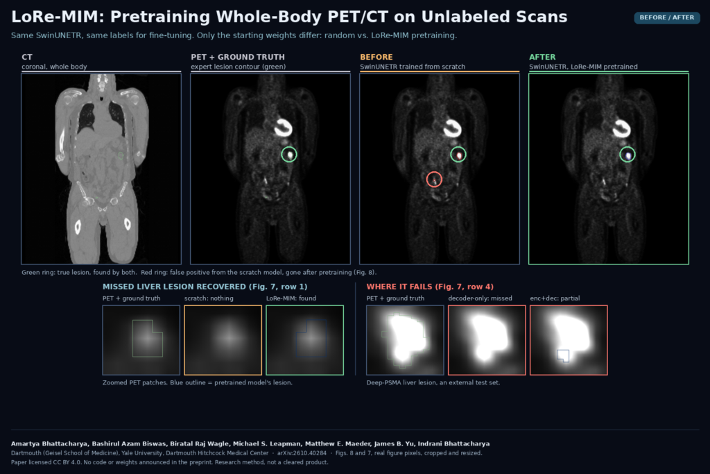
Before and after panel from Figures 8 and 7 of arXiv:2610.40284 by Amartya Bhattacharya, Bashirul Azam Biswas and colleagues (Dartmouth, Yale and Dartmouth Hitchcock Medical Center): coronal whole-body CT and PET with the ground-truth lesion, a SwinUNETR trained from scratch that adds a false positive, and the LoRe-MIM pretrained model that keeps the lesion and drops the false positive, plus zoomed PET patches of a recovered liver lesion and a Deep-PSMA liver lesion that the pretrained models miss or only partly segment.

A masked-image pretext that swaps PET and CT blocks helps a SwinUNETR find liver and bone lesions and drop false calls.

Self-Training Coronary Vessel Masks With Pre-Contrast Frames
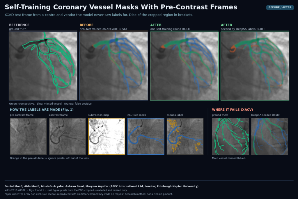
Before and after panel from Figures 2 and 1 of arXiv:2610.40302 by Danial Moafi, Aida Moafi, Mostafa Aryafar, Ashkan Sami and Maryam Aryafar (APEC International Ltd and Edinburgh Napier University): an XCAD coronary angiogram with ground truth, the ARCADE-trained nnU-Net, the self-trained model and the DeepSA-seeded model, a strip showing the pre-contrast frame, contrast frame, subtraction map, seed mask and pseudo-label, and an XACV frame where the main vessel is missed.

An nnU-Net labels a new hospital’s angiograms, and the frame before contrast arrives flags the branches it missed.

RepConTUR: Report-Prompted 3D Tumor Contours for Abdominal CT
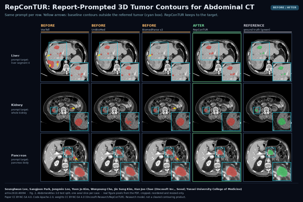
Before and after panel from Figure 2 of arXiv:2610.40094 by Seunghwan Lee, Sangjoon Park, Jongmin Lee, Yoon Jo Kim, Wonyoung Cho, Jin Sung Kim and Han Joo Chae (Oncosoft Inc. and Yonsei University College of Medicine): axial abdominal CT slices for liver segment 4, kidney and pancreas body tumor prompts, comparing VoxTell, UniBioMed and BiomedParse v2 masks with yellow arrows on off-target contours, the RepConTUR mask and the ground truth.

The report names the liver segment or pancreas region, and a 3D model contours that tumor with fewer stray masks elsewhere.

MRI-Free Amyloid PET Quantification With Calibrated Error Bars
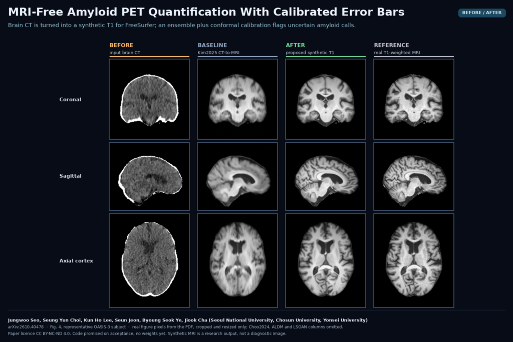
Before and after panel from Figure 4 of arXiv:2610.40478 by Jungwoo Seo, Seung Yun Choi, Kun Ho Lee, Seun Jeon, Byoung Seok Ye and Jiook Cha (Seoul National University, Chosun University, Yonsei University): coronal, sagittal and axial slices of an OASIS-3 subject showing the input brain CT, the Kim2025 synthetic T1, the proposed synthetic T1 and the real T1-weighted MRI.

Brain CT becomes a synthetic T1 for FreeSurfer, and an ensemble with conformal calibration flags the amyloid calls that need an MRI.

Pancreatic Duct Width From 3D MRCP, With a Review Flag
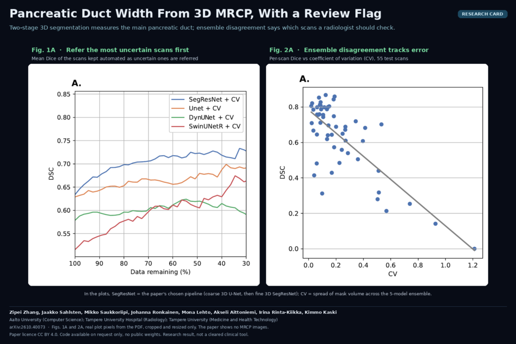
Research card with Figures 1A and 2A of arXiv:2610.40073 by Zipei Zhang, Jaakko Sahlsten, Mikko Saukkoriipi, Johanna Ronkainen, Mona Lehto, Akseli Aittoniemi, Irina Rinta-Kiikka and Kimmo Kaski (Aalto University, Tampere University Hospital, Tampere University): mean Dice of automated scans as uncertain cases are referred for four pipelines, and per-scan Dice against ensemble coefficient of variation.

A two-stage 3D model segments the main pancreatic duct on MRCP, and ensemble disagreement marks the scans a radiologist should check.

Spine MRI-to-CT Synthesis With Vertebra-Matched Pseudo-Pairs
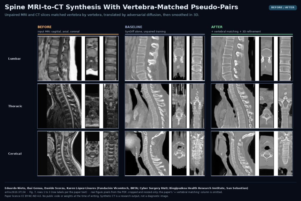
Figure 7 of arXiv:2610.37134 by Eduardo Nieto, Ibai Genua, Davide Scorza and Karen Lopez-Linares (Vicomtech, Cyber Surgery, Biogipuzkoa): lumbar, thoracic and cervical spine MRI in sagittal, axial and coronal views, the synthetic CT from the SynDiff baseline, and the synthetic CT from the full pipeline with vertebral matching and 3D refinement.

Unpaired MRI and CT slices are matched vertebra by vertebra, translated with adversarial diffusion, then cleaned up in 3D.

← Previous
Next →